
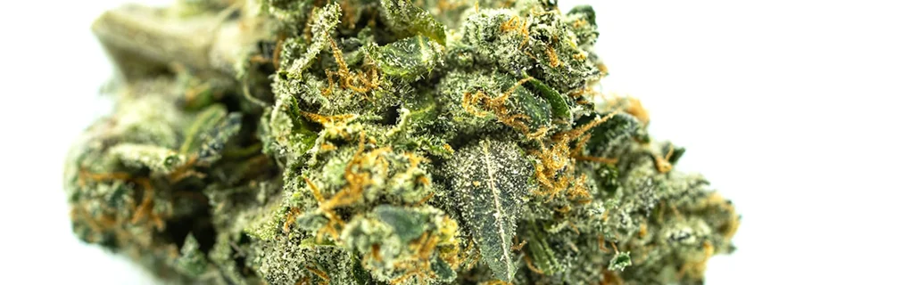
Cannabis pathogen testing vs. visual diagnosis
Every experienced cannabis grower has made this mistake: seeing a problem, attributing it to the most plausible environmental cause, adjusting the environment, and not seeing improvement. The missed diagnosis is usually not the grower’s fault — cannabis pathogens and environmental stressors produce overlapping symptoms, and without molecular testing, there is genuinely no reliable way to tell them apart.
The Core Problem
Visual diagnosis tells you what a symptom looks like. It cannot tell you what is causing it.
The most economically damaging pathogens — HLVd, CCV, and early-stage Fusarium — produce no visible symptoms during the period when they do the most damage. By the time visual signs appear, the infection has already spread.
The four ways visual diagnosis misleads growers
The fundamental problem is that plant symptoms are not pathogen-specific. A nitrogen-deficient plant and an HLVd-infected plant can look indistinguishable. A plant wilting from overwatering and a plant wilting from Pythium root rot look the same until you pull the roots.
Asymptomatic infections
HLVd and CCV produce no visible symptoms during early infection — which can last weeks to months. The plant looks healthy. It is not. By the time any visual signal appears (if it appears at all), the infection has been replicating and spreading through your propagation system. Visual diagnosis cannot detect what is invisible.
Symptom overlap
The symptoms HLVd eventually produces — tight internodes, slightly pale leaves, reduced resin — are identical to symptoms produced by light stress, temperature fluctuation, nutrient imbalance, root restriction, and genetics. In a commercial environment with multiple variables in play, there is no reliable way to attribute these symptoms to a pathogen by sight.
Confirmation bias
When a grower sees wilting in a plant that was recently watered, the most available explanation is overwatering. When they see tight internodes, the most available explanation is the light schedule. These explanations are often correct — which is exactly why they are so effective at masking the less common but more serious explanation: pathogen infection.
Delayed detection compounds losses
The practical consequence of relying on visual diagnosis is that detection happens late — at harvest, when product quality disappoints, or when symptoms become dramatic enough to investigate. By that point, the infection has already distributed through propagation. The financial damage is done. Molecular testing can only confirm what happened.
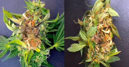
Every symptom, multiple possible causes
The table below maps common cannabis visual symptoms to their most frequent misdiagnoses and their actual range of possible causes. The pattern is consistent: every symptom that a pathogen produces is also produced by at least one environmental stressor.
| Symptom | Visual Diagnosis Says | Actual Possible Causes |
|---|---|---|
| Tight internodes | Light stress or genetics | HLVd, genetics, light intensity, root restriction |
| Pale or small leaves | Nitrogen deficiency | HLVd, CCV, nutrient deficiency, pH imbalance, root disease |
| Wilting despite wet medium | Overwatering or root rot | Pythium myriotylum, Fusarium oxysporum, HLVd, overwatering |
| Yellowing lower leaves | Nitrogen deficiency or nenescence | Fusarium solani, Pythium myriotylum, BCTV, LCV, HLVd, normal nenescence |
| Loose bud structure | Poor genetics, humidity, harvest timing | HLVd, CCV, genetics, humidity, nutrient imbalance |
| Reduced trichome density | Late harvest, genetics, or stress | HLVd, CCV, genetics, light quality, harvest timing |
| Gray mold on flowers ✓ Reliable | Botrytis — correct | Botrytis cinerea (visual diagnosis reliable for this one) |
| Stem lesions or rot | Mechanical damage or Fusarium | Fusarium oxysporum, Fusarium solani, physical damage |
The table illustrates the diagnostic problem precisely. Virtually every symptom that matters has multiple possible causes — only one of which requires quarantine, pathogen removal, and possibly tissue culture recovery. Visual inspection alone cannot distinguish between them. The one exception is Botrytis cinerea gray mold, which has a characteristic appearance that makes visual identification reasonably reliable. For everything else, molecular confirmation is the only reliable answer.
When visual diagnosis is appropiate and when it's not
Visual diagnosis is not useless. It is appropriate for certain decisions and completely inadequate for others.
The distinction matters for how you allocate testing resources.
Where visual diagnosis IS appropiate
- Identifying obvious mechanical damage, physical defects, or environmental conditions that have an unambiguous visual signature
- Confirming Botrytis cinerea (gray mold) with white-gray mycelial growth distinctive enough to identify visually
- Triaging which plants to test first — symptomatic plants may warrant priority testing, even though asymptomatic plants also need screening
- Monitoring plant response after a confirmed diagnosis and treatment decision — visual assessment of recovery or decline when the cause is already known
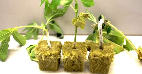
- Identifying damping-off in seedlings and clones: rapid collapse at the soil line, water-soaked or necrotic tissue, white mycelial growth. Useful for immediate triage, but lab testing is required to confirm the causal organism
Where visual diagnosis is NOT appropriate
- Clearing new genetics as clean before entering the mother room — HLVd has no visible early-stage symptoms
- Ruling out pathogen infection when yield or quality falls below expectation
- Making quarantine or removal decisions about mother plants — only molecular results justify action
- Clearing plants after a positive result and remediation — only a subsequent negative molecular test confirms clearance. For HLVd, once confirmed, the plant must be removed
- Any decision that affects propagation stock — consequences of a missed positive are too high to rely on visual assessment
- Distinguishing between pathogens and abiotic stress — nutrient issues, irrigation problems, and environmental stress mimic pathogen symptoms
- Visual symptoms do not correlate reliably with pathogen load — plants with mild symptoms may still carry significant infection
- Visual health does not indicate pathogen status — long-lived mother plants require routine molecular testing regardless of appearance
The exception: Botrytis cinerea (gray mold)
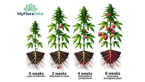
Why location and tissue type affect detection
HLVd is not uniformly distributed throughout the plant, particularly during early infection. Research shows the viroid can be detected in roots and young tissues before it is consistently detectable in mature leaves.
Over time, HLVd spreads systemically throughout the plant. But early on, uneven distribution creates a sampling problem. Testing a single leaf or a single location can miss early infections, even when the plant is already infected and capable of spreading the viroid.
For reliable detection, sample:
Young growth tissue
New leaves and actively growing shoots where HLVd is often detectable early.
Mature leaves
Fan leaves from the mid-plant — commonly sampled but not always the earliest to show detectable load.
Root-associated material
Roots can carry detectable viroid load before leaves do — sample where possible.
Multiple plants
Testing multiple plants within a population further reduces the risk of false negatives.
The clearest statement of when molecular testing is required: any decision about whether a plant should be propagated from requires a molecular test result. Visual assessment is not sufficient to make that call.
Reactive vs. proactive testing
Reactive testing — ordering a test after a visible problem appears — only confirms a diagnosis after the damage is done. Proactive testing through scheduled molecular screening before any symptoms appear catches infection during the window when quarantine actually prevents spread.
Triggered by visible symptoms — after the window has closed
By the time symptoms appear, the infection has already spread through tools, water, and propagation systems. The test only confirms what happened. The damage is already embedded in the pipeline.
Scheduled screening before any symptoms appear
Quarterly pathogen panel testing for mother rooms, plus screening of all incoming genetics before integration. Catches infection while still contained to a limited number of plants.
The asymptomatic window is where the damage happens
After HLVd enters a mother plant, it begins replicating immediately. For the first several weeks, there are no visual signals. During this period, if cutting sessions continue without sterilization between plants, the viroid can be distributed to every plant that shares tools with the infected mother.
Spread is not limited to tools. In propagation systems — especially those using shared water sources — HLVd can move through root contact, recirculating irrigation, and contaminated solution. Clones rooted in the same tray, cloner, or hydroponic system can be exposed simultaneously, even without direct handling.
If cuttings from an infected plant have already been rooted and transferred to vegetative, those plants carry the infection forward into production.
At that point, the issue is no longer a single plant — it is embedded in the propagation pipeline.
The proactive testing window is the asymptomatic phase. MyFloraDNA recommends quarterly pathogen panel testing for the mother room, along with screening of all incoming genetics before integration into existing rooms.
The cost difference between proactive and reactive
Proactive Cost
$18 / sample Scheduled panel per mother plant, quarterly. Multiplex qPCR covers 3 pathogens simultaneously.
Reactive Consequence
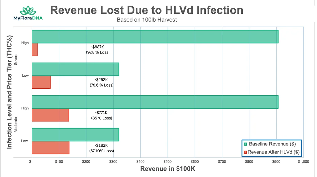
$771,000+ Revenue loss from undetected moderate HLVd in a 100 lb high-tier harvest — plus removal, sanitation, and genetic recovery costs.
Proactive testing at any realistic cadence costs orders of magnitude less than the revenue lost to a single undetected infection. See the full economic model in the Economic Impact of HLVd cluster guide.
Building a two-Signal diagnostic workflow
An effective diagnostic workflow integrates visual monitoring with scheduled molecular testing and a clear decision tree for what happens when each type of signal fires.
Signal 1: Scheduled molecular testing
Regardless of what plants look like, every mother plant is screened on a quarterly schedule. These tests run whether anything looks wrong or not.
- Quarterly mother room baseline (full room)
- Incoming genetics screened before integration
- Pre- and post-tissue culture material at each checkpoint
- Runs on schedule — not triggered by appearance
Signal 2: Visual monitoring (additional layer)
When a plant shows symptoms that could indicate pathogen involvement, an out-of-cycle test with a symptom-specific panel is triggered.
- Unexplained wilting → Wilting Panel™
- Unusual yellowing → Yellowing Panel™
- Reduced vigor → General Decline Panel™
- Adds an additional trigger layer — does not replace scheduled testing
For the specific testing cadence, quarantine procedures, and staff protocols that operationalize this approach, see the Cannabis Mother Room Biosecurity Guide. For the multiplex qPCR technology that makes proactive testing cost-effective, see What Is Multiplex qPCR in Cannabis Testing?
Don’t wait for symptoms. Test on schedule.
Submit a sample and get quantitative pathogen data via MyFloraCLOUD in 72 hours or less.
Frequently Asked Questions
Botrytis cinerea (gray mold) has a characteristic appearance that makes visual identification reasonablyreliable in most cases. Severe late-stage Fusarium with obvious vascular discoloration can be visually suspected, though molecular confirmation is still required before quarantine decisions. HLVd, CCV, Pythium, and early-stage Fusarium cannot be reliably diagnosed visually.
Yes, if it is in the mother room or is a candidate for propagation. HLVd can be present at significant load with no visible symptoms at all. A completely healthy-looking mother plant can distribute HLVd to every cutting it produces during an undetected infection.
Cannabis plants invest in secondary metabolite production (cannabinoids, terpenes) as a response to stress and as part of reproductive development. HLVd disrupts the gene expression pathways involved in this process without necessarily disrupting primary growth. The plant grows, flowers, and appears to develop normally — but produces significantly less of the compounds that determine its market value.
No. The yield and potency suppression caused by HLVd is a biological effect of viroid infection on gene expression rather than a response to suboptimal conditions. Improving light, nutrients, or environment will not restore cannabinoid content in an infected plant. The only solution is removal and replacement with clean genetics.
Treat it the same as a symptomatic positive: quarantine immediately, stop cutting, trace prior cuttings, re-test adjacent plants. The absence of visible symptoms does not change the infection status or the quarantine requirement. In some ways, an asymptomatic positive is the more dangerous result — it confirms that visual monitoring would never have caught this plant.
Resources
Related Guides
Diagnostics, biosecurity, and remediation guidance